CASE REPORT | https://doi.org/10.5005/jp-journals-10077-3077 |
An Unusual Foreign Object Encircling Primary Teeth: A Case Report
Department of Pediatric Dentistry, Dr NTR University of Health Sciences, Bhimavaram, Andhra Pradesh, India
Corresponding Author: Madhuri Vegesna, Department of Pediatric Dentistry, Dr NTR University of Health Sciences, Bhimavaram, Andhra Pradesh, India, Phone: +91 9491088088, e-mail: madhuvegesna@yahoo.co.in
How to cite this article Madhuri V. An Unusual Foreign Object Encircling Primary Teeth: A Case Report. J South Asian Assoc Pediatr Dent 2021;4(2):128–130.
Source of support: Nil
Conflict of interest: None
ABSTRACT
Fortuitous placement of objects into the mouth and nibbling while playing is common among infants and toddlers. Most of the time, it is unnoticed until the child complains of pain or discomfort. Sometimes, the inadvertent gnawing of these tiny bits and pieces may injure the oral tissues leading to infections if not intervened at an early stage. It is quite challenging to remove such foreign objects and preserve the involved teeth, especially in young children. This case report highlights one such incident in a 15-month-old child which cost him his lower two central incisors.
Keywords: Foreign object, Primary teeth, Plastic ring.
INTRODUCTION
Children have a common tendency to insert foreign objects in their oral cavity specifically those that are readily available and easily inserted such as small toys, pins, coins, marbles, screws, toothpicks, stapler pins, and pencil leads.1,2 Accidental ingestion or aspiration of these objects may cause severe discomfort and can pose a serious threat when associated with pain, swelling, and trauma.1 The risk of impaction of these foreign objects is high especially in teeth with open pulp chambers, large carious exposure, or incomplete root canal procedures.3 Ingested metal objects may react with body fluids and acids generating toxic chemicals. There is also a probable risk of tiny items being aspirated leading to airway obstruction. Aspiration of foreign bodies occurs mostly in kids younger than 5 years, with 65% of deaths involving infants younger than 1 year.4,5 Several cases have been reported wherein such objects have been accidentally lodged into the tooth especially in teeth with large carious lesions. However, a foreign object lodged around the tooth is fairly uncommon. The following is a case report describing the retrieval of a foreign object encircling the teeth of a child.
CASE DESCRIPTION
A 15-month-old child accompanied by his parents reported to a private dental clinic in Bhimavaram. The parents complained of a hard structure attached to the tooth due to which the child had difficulty in eating and brushing because of bleeding of the gingiva. The parents reported that the teeth had started erupting two months back and immediately this hard structure was seen around the teeth. The parents had presumed the foreign object to be a growing part of the tooth.
Intraoral examination revealed a thick hard structure enveloping around the two lower central incisors covering the cervical 1/3rd of the crown and also the root, measuring about 0.5 × 0.5 × 0.4 cm, smooth in surface, cream in color, and hard in consistency (Fig. 1). The gingiva around the hard structure and tooth looked inflamed. Both the central incisors and the hard structure were mobile. Specimen radiograph revealed a radiopaque halo-like structure enveloping the two lower central incisors and loss of periodontium around them. The hyperdense ring-like structure exhibited no difference in radiodensity when compared with enamel (Fig. 2).
Since the child had discomfort during eating and brushing it was decided to surgically remove the hard structure. Under local anesthesia, the hard structure enveloping the teeth was first removed following which both the central incisors were removed as there was grade III mobility due to loss of periodontium (Fig. 3).

Fig. 1: Intraoral photograph shows the plastic ring around the lower central incisors
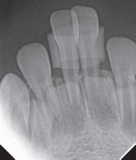
Fig. 2: Intraoral periapical radiograph shows the radiodense ring around the teeth and loss of periodontium

Fig. 3: Extracted teeth and plastic ring

Fig. 4: Polarized microscope image of the cut section of polyvinyl chloride tubing depicts polarization
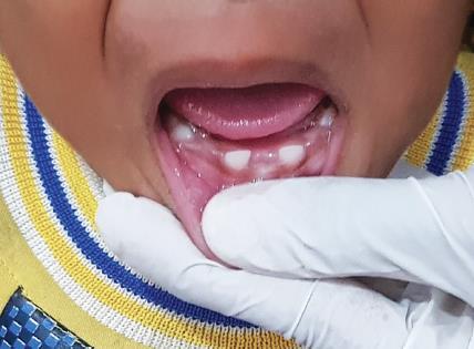
Fig. 5: Intraoral photograph shows healed sockets after 1-week follow-up
The specimens were sent for histopathological analysis. When examined under stereomicroscope, the surface of the foreign body showed a corrugated appearance resembling that of bone but on palpation the ring was compressible. The cut surface of the 1 mm section examined under polarized light showed a multicolored surface appearance which was changing constantly while adjusting the fine adjustment and also by light intensity. The 10 μm and 20 μm sections exhibited structureless masses of plastic material (Fig. 4). Based on the clinical and histopathological report, the foreign object was confirmed to be a plastic/polyvinyl chloride (PVC) structure. The child would have accidentally placed the plastic ring around his teeth while playing. The parents have failed to notice it until the child had discomfort.
After a 7-day follow-up, the extraction sockets and gingiva around the hard structure have healed (Fig. 5). The parents reported the child was able to eat and brush without discomfort. However, a space maintainer could not be given at this stage because there were no teeth to support it.
DISCUSSION
Foreign objects inserted in the tooth vary from radiolucent objects like toothbrush bristle and wooden toothpicks to radiopaque materials such as pins, beads, needles, screws, and pencil leads. The detection of such foreign bodies in the teeth is an unusual situation that often is diagnosed accidentally.1,6 In the present case, the foreign object retrieved was a plastic ring that was embedded around the lower two central incisors. The child must have accidentally pushed it around the teeth. Unfortunately, the parents were unaware and they had assumed the teeth were growing abnormally.
Both hard and soft tissues lesions may occur as a consequence of child′s habit of placing foreign items into their mouth.7 The parents reported to the dentist only when the child had complained of pain and bleeding gums. The teeth had exhibited severe mobility due to which both the lower central incisors had to be extracted. Continuous impingement of the plastic ring around the tooth for 2 months has lead to loss of periodontium resulting in the mobility of the lower central incisors. Objects embedded within the periodontium are a potential source of infection resulting in bleeding, edema, and abscess formation.8 Foreign bodies implanted in deciduous dentition can perforate the floor of the pulp chamber and possibly interfere with developing permanent tooth germ resulting in odontoma, impaction, or dilaceration.5 Any insult to the tooth during odontogenesis can affect the tooth bud completely or may cause disorganization of the tooth germ.8 Such foreign objects may also hinder the eruption of adjacent or underlying primary or permanent teeth.
Retrieval of foreign bodies from the teeth in children is a demanding aspect of pediatric dentistry. These objects may be impregnated in the cavities of the broken or fractured teeth and the root canals of the teeth with open pulp chambers, gums, tongue, the floor of the mouth, tonsillar fossa and crypts, and jawbones.9 Passi and Sharma cited a series of cases in which foreign bodies such as a stapler pin, tip of the metallic compass, broken sewing needle, and a copper strip were impregnated in the tooth.2 Kanika reported a case of an 8-year-old child where a ball pin was lodged between mandibular deciduous molars.10 The treatment of a foreign body impaction in a tooth is influenced by the site, ease of access, stage of tooth development, restorability of the tooth, the child’s age, and level of cooperation.8
Clinical presentation may be in the form of a simple abscess or occasionally mimicking other pathologies such as granulomas, tumors, etc.9 Comprehensive case history and clinical examination is vital to determine the etiology, size, site, and type of the foreign material.11 In this case, the child could not give any history as he is too young to speak. Azodo et al. published a similar case of a 6-year-old child where a piece of the inner tube of biro was embedded on 72. The child was reported to have initially denied chewing the biro for fear of being punished.12
The lodged foci of infection have to be eliminated at the earliest to avoid complications. Also, there may be alarming consequences, such as aspiration of the foreign body. The pushing of foreign bodies into the maxillary sinus through the root canals has lead to chronic maxillary sinusitis of a dental origin as reported by Costa.13 Timely diagnosis can prevent complications such as ingestion and damage to the teeth.14 Timely removal of the plastic ring, in this case, would have aided in preserving the two central incisors. Parents need to be warned to keep small objects away from children to avoid such accidents. Anticipatory guidance needs to be provided to parents to avoid emergencies and aid in timely intervention.
REFERENCES
1. Holla G, Baliga S, Yeluri R, et al. Unusual objects in the root canal of deciduous teeth: a report of two cases. Contemp Clin Dent 2010;1(4):246–248. DOI: 10.4103/0976-237X.76393.
2. Passi S, Sharma N. Unusual foreign bodies in the orofacial region. Case Rep Dent 2012;2012:191873. DOI: 10.1155/2012/191873.
3. Khandelwal D, Kalra N, Tyagi R, et al. Accidental diagnosis of a foreign body embedded in maxillary anterior tooth. J Sci Soc 2019;46(3):103–105. DOI: 10.4103/jss.JSS_29_19.
4. Lakhani B, Garg S, Saraf BG, et al. Self-insertion of foreign objects in teeth. Int J Clin Pediatr Dent 2019;12(2):145–149. DOI: 10.5005/jp-journals-10005-1595.
5. Chandran N, Sudeep CB, Johny J, et al. Foreign body in maxillary deciduous first molar – a case report. Int J Contemp Med Res 2020;7(6):F4–F5. DOI: 10.21276/ijcmr.2020.7.6.8.
6. Setty JV, Srinivasan I. Beads in the tooth. Int J Clin Pediatr Dent 2011;4(2):163–165. DOI: 10.5005/jp-journals-10005-1103.
7. Aduri R, Reddy RE, Kiran K. Foreign objects in teeth: retrieval and management. J Indian Soc Pedod Prev Dent 2009;27(3):179–183. DOI: 10.4103/0970-4388.57100.
8. Katge F, Mithiborwala S, Pammi T. Incidental radiographic discovery of a screw in a primary molar: an unusual case report in a 6 year old child. Case Rep Dent 2013;2013:296425. DOI: 10.1155/2013/296425.
9. Ajike SO. Impacted foreign bodies in dentistry. J West Afr Coll Surg 2015;5(3):x–xi.
10. Dhull KS, Acharya S, Ray P, et al. Foreign body in root canals of two adjacent deciduous molars: a case report. Int J Clin Pediatr Dent 2013;6(1):38–39. DOI: 10.5005/jp-journals-10005-1184.
11. Mahesh C, Animireddy D, Mettu S, et al. An unexpected encounter with foreign body in the primary teeth and its management. Int J App Basic Med Res 2018;8(3):181–183. DOI: 10.4103/ijabmr.IJABMR_264_17.
12. Azodo CC, Erhabor P, Chukwumah NM, et al. Intraoral foreign body: a case report and review of literature. Indian J Multidiscip Dent 2015;5:97–99.
13. Gujjar KR, Algali G, Omar SM, et al. Foreign bodies in primary molars: a report of 2 cases. J Dent Child (Chic) 2012;79(1):40–43.
14. Kanumuri PK, Gantha SN, Animireddy D, et al. Unusual foreign body in primary tooth. BMJ Case Rep 2016;2016:bcr2016216326. DOI: 10.1136/bcr-2016-216326.
________________________
© The Author(s). 2021 Open Access This article is distributed under the terms of the Creative Commons Attribution 4.0 International License (https://creativecommons.org/licenses/by-nc/4.0/), which permits unrestricted use, distribution, and non-commercial reproduction in any medium, provided you give appropriate credit to the original author(s) and the source, provide a link to the Creative Commons license, and indicate if changes were made. The Creative Commons Public Domain Dedication waiver (http://creativecommons.org/publicdomain/zero/1.0/) applies to the data made available in this article, unless otherwise stated.